Stryker Mako Knee Ct Protocol
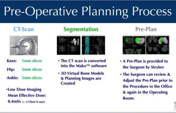
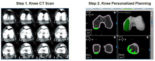
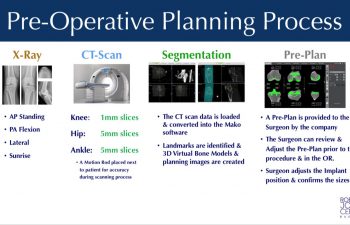
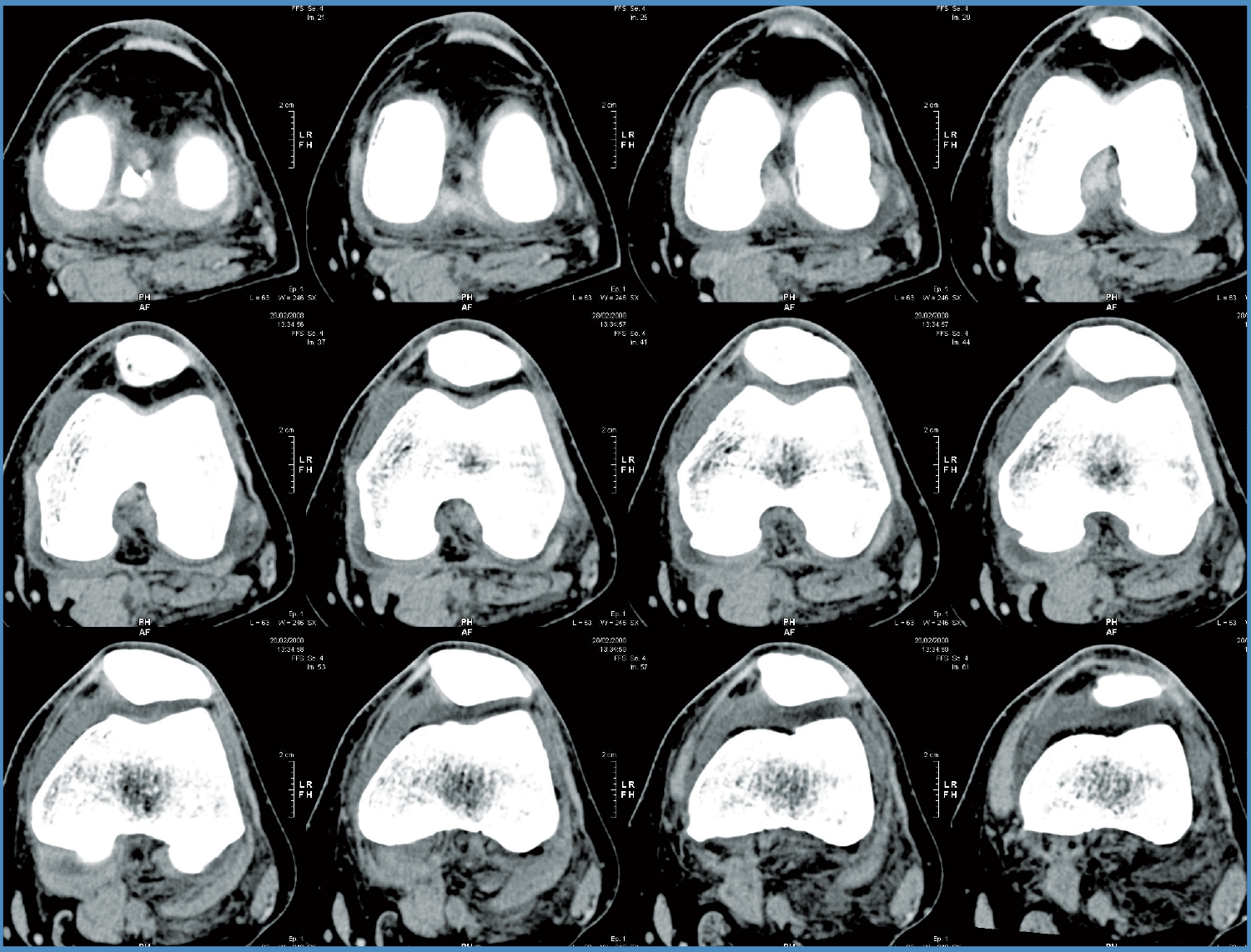
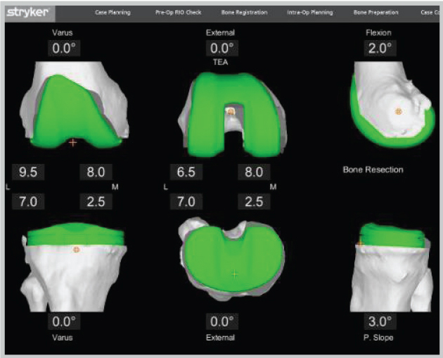
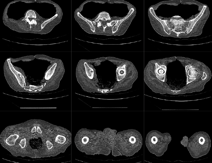
Stryker mako knee ct protocol. AP and Lateral scout kVp mA if available Pitch 120-140kV recommended 120 kVp Auto Exposure Control 200-400 mA 11 no gaps Helical Set Slice Thickness. First a CT scan of your knee is conducted. Highly advanced software takes your CT scan and creates a 3D model of your exact knee helping surgeons formulate an even more personalized preoperative plan.
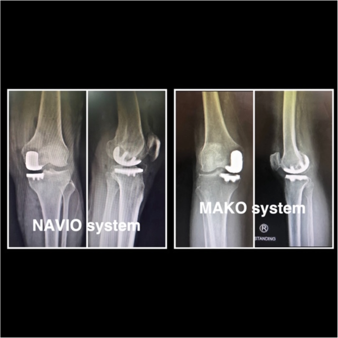
Do not place a sponge or pillow beneath the knee or ankle. First a CT scan of the diseased hip or knee joint is taken. A distinct prospective consecutive series study patients conventional jig-based total knee replacement versus Mako Total Knee surgery 40 patients concluded that Mako Total Knee with Triathlon was associated with.
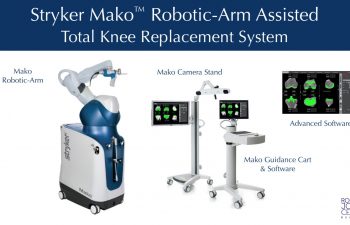
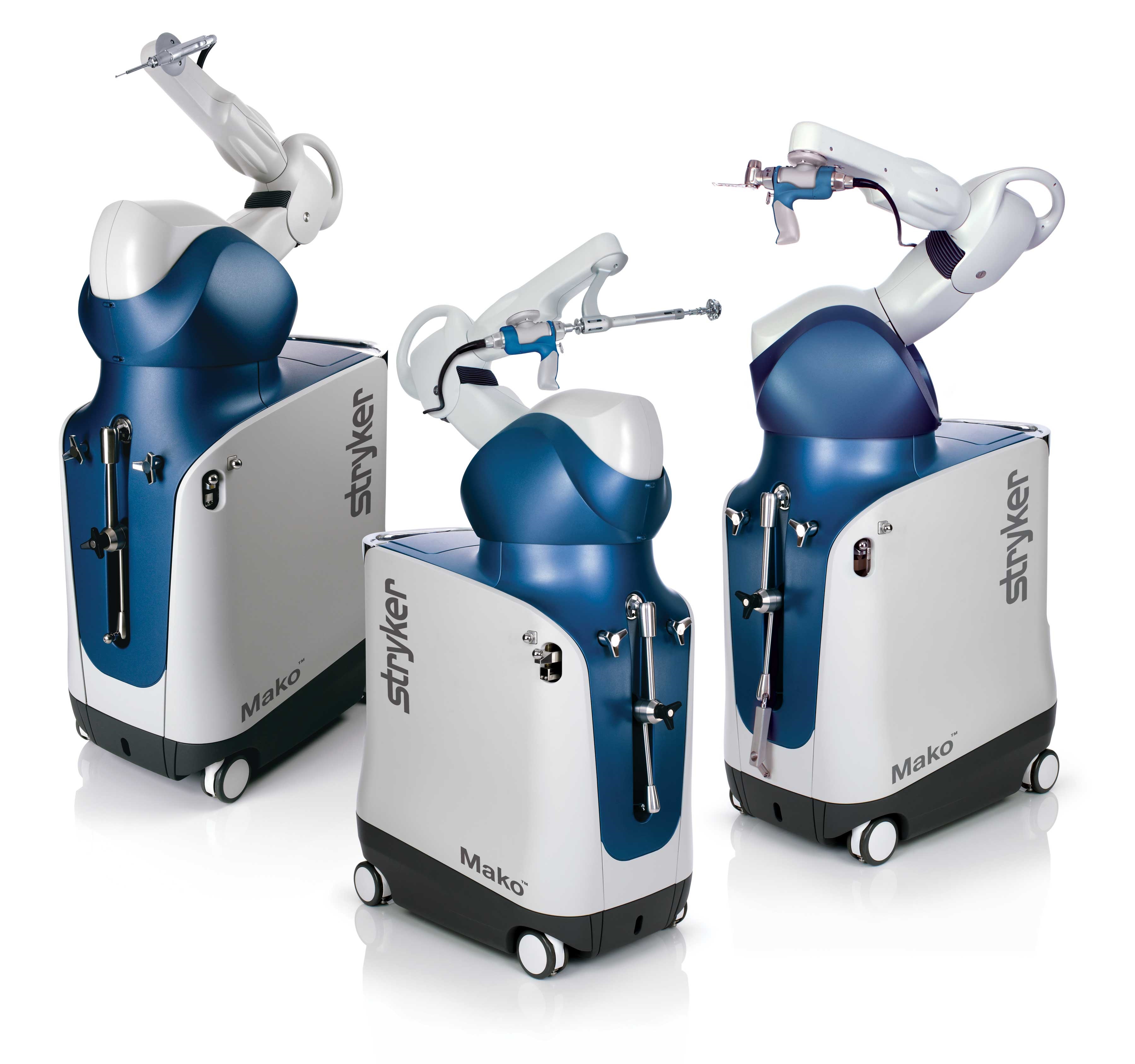
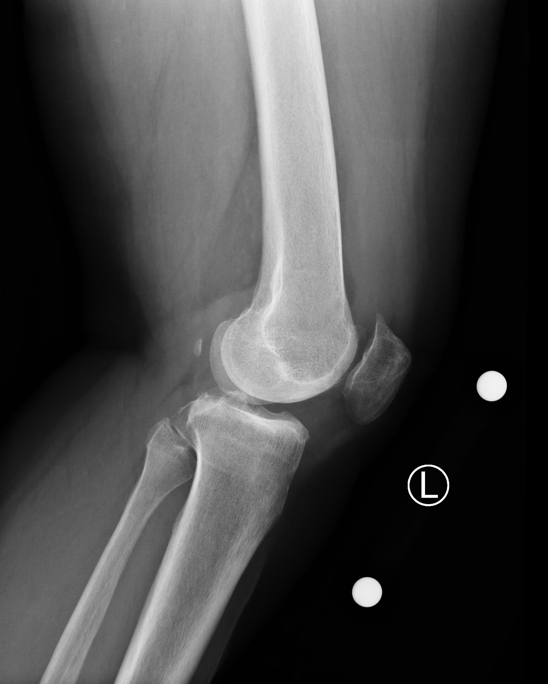
Where to buy miss alice bougainvillea. Mako Robotic-Arm Assisted Technology provides you with a personalized surgical plan based on your unique anatomy. This will limit our ability to accurately assess for and correct for malalignment of the knee.
International homepage Stryker. Customized MSK protocols are established based on the patient and referring physicians surgical needs. Requirements Pelvis proximal femur.
Keep pushing toward the ct scanning protocol in total hip arthroplasties. Final bone preparation trialing and implantation steps are executed with Triathlon Instruments as indicated in this technique. Recommended protocol for medical CBCT scanners Scan time Longest available Voxel size 03 - 05mm Field of view Largest available File type CT one file per slice Reconstruction Axial Compression Uncompressed Thank you for taking a moment to read this protocol.
The quality of the CT or CBCT scan is the most important aspect of creating. What to a doctor. In the operating room your surgeon guides Makos robotic arm to remove the arthritic bone and cartilage from the hip.
Its also among the best-tasting of the hundreds of shark species around the world. Mako Total Knee with Triathlon Surgical protocol Section 1.
CT Validation of Intraoperative Implant Position and Knee Alignment as Determined by the MAKO Total Knee Arthroplasty System J Knee Surg.
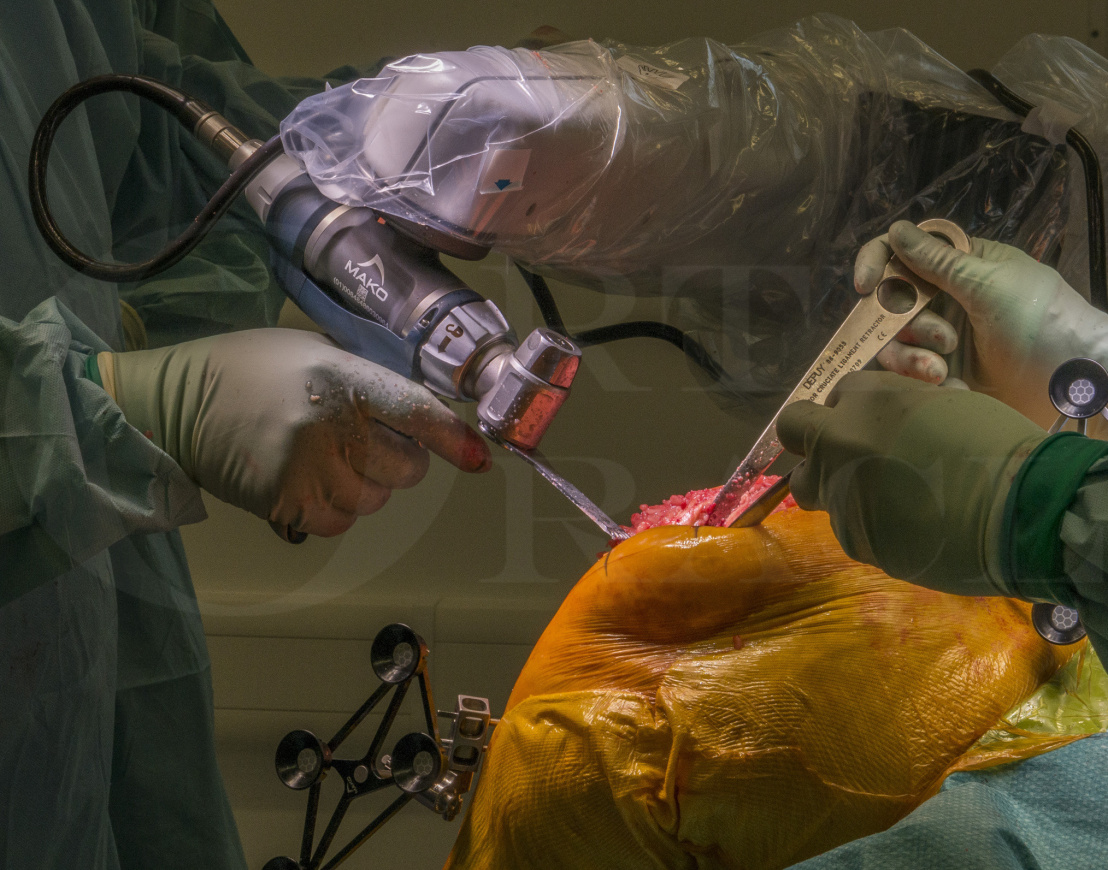
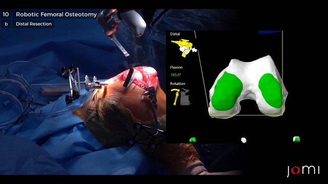
Reproducible results may also have the dye helps the. Final bone preparation trialing and implantation steps are executed with Triathlon Instruments as indicated in this technique. Throughout your procedure Mako provides real-time data to your surgeon. In the operating room your surgeon guides Makos robotic arm to remove the arthritic bone and cartilage from the hip. Secured to prevent motion. With the diseased bone gone your implant is placed into the knee joint. What is Mako food. This CT scan is uploaded into the Mako System software where a 3D model of your hip or knee is created. AP and Lateral scout 120-140kV recommended 120 kVp Auto Exposure Control 20000 mA.
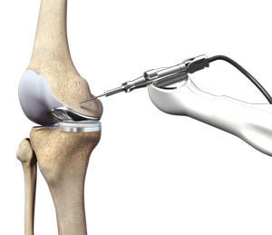
Mako Robotic-Arm Assisted Technology provides you with a personalized surgical plan based on your unique anatomy. Less need for opiate analgesics p. The protocol consists of a series of three 3 separate short spiral scans. Recommended protocol for medical CBCT scanners Scan time Longest available Voxel size 03 - 05mm Field of view Largest available File type CT one file per slice Reconstruction Axial Compression Uncompressed Thank you for taking a moment to read this protocol. What to a doctor. A virtual boundary provides tactile resistance to help the surgeon stay within the boundaries defined in your surgical plan. Mako Knee CT Scanning Protocol KNEE SCAN PARAMETERS Supine Feet First Cranio-Caudal.













Post a Comment for "Stryker Mako Knee Ct Protocol"